|
1. REU-Site: Wellman-HST Summer Institute for Biomedical Optics In collaboration with the Harvard-MIT Health Sciences and Technology (HST), the Wellman Center has run the Summer Institute for Biomedical Optics since 2002.
This program provides undergraduate student participants with research experience in the field of biomedical optics. The program objective is to inspire talented students to pursue advanced research, education, and careers in science and engineering. Faculty mentors offer interdisciplinary cutting-edge research projects in diverse, yet cohesive, themes in biomedical optics.
8-10 undergraduate students admitted each summer pursue full-time laboratory research for nine weeks, working in one of the laboratories at the Wellman Center and MIT. In addition to research, students attend lectures and research seminars, as well as professional development workshops on scientific writing, presentation and research ethics. All student participants are accommodated in the dormitory, receive weekly stipends, and participate in various peer group activities, making their summer experience go much beyond what could be provided by a typical summer research experience in a single lab.
Research experience in this program is focused on engineering from discovery of new transformative approaches to development of cutting-edge technologies. Innovation in biomedical optics requires understanding of physical and engineering principles, as well as biological and medical insights, to define important challenges and to understand how new technologies must perform. This program takes a coherent and interdisciplinary approach to introducing students to the full spectrum of biomedical optics and help to realize the promise of biomedical optics by attracting talented students to pursue careers in this area at the graduate level and beyond.
Details and an application form can be obtained at the MIT-HST web site.
Students must be a sophomore, junior, or senior undergraduate at the time of the program and must also be a United States Citizen or have permanent residence status.
Benefits: · $5,000 stipend · Housing · Travel support ~$400 · Opportunity to present at NSF conference · Mentor-mentee relationship
2. MGH-KAIST Summer Internship Program
3. MGH-Tokyo Summer Training Program
4. MGH-China Summer Exchange Program
— Tongji Medical School — Jiao Tong Medical School
General Information
Inquires to: BioOpticsSummerInstitute@mgh.harvard.edu
Organization Structure Executive Director: Prof S. H. Andy Yun Deputy Director: Dr. Walfre Franco Program coordinator: Jonathan Lawson Registration: Deana Marzullo Mentors: All Wellman Faculty
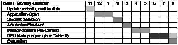
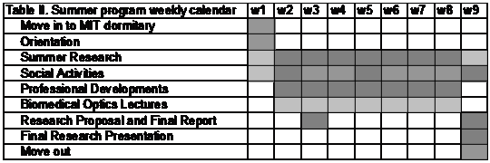
Timetable The duration of the summer program is nine weeks, typically lasting from the second week of June until the first week of August. The schedule is optimized to give admitted students sufficient time to prepare before the program starts as well as sufficient time to prepare for the fall semester after the program ends. The detailed schedule is as follows:
The summer schedule (from the second week of June to the first week of August) is as follows:
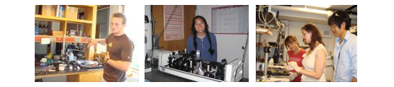
Summer Research The students pursue full-time laboratory research during the nine-week period. Each student works in direct connection with faculty mentors as well as graduate students and post-doctoral fellows in the lab. At least one senior student or postdoc is assigned to supervise each student on a daily basis. Each student participates in one summer project with a clearly defined and attainable objective. Typically, summer projects are designed such that students can work with a high degree of independence while leveraging from other ongoing research in the host lab. In general, a student spends approximately 50% of his or her time in the lab and the other 50% in the office writing, reading, and analyzing data.
By conducting hands-on experiments, students learn about cutting-edge engineering and technologies applicable to biomedical problems. Equally important, they learn about multidisciplinary research, how it is organized and conducted, and how to communicate with members of a research team with very diverse backgrounds. Students also learn how to think about their problem creatively and how to proactively pursue their research objectives by seeking help from experts, trying out new approaches, developing skills to delve deeper into problems, documenting their efforts, writing up their work for papers and patents, and other strategies. Summer students prepare weekly summaries of their research progress for discussion with their mentor and other lab members during group meetings.
Summer Research Topics
The students are engaged in full-time laboratory research during the 10-week period. Each student works directly with a faculty mentor as well as graduate students and post-doctoral fellows in the lab. At least one senior graduate student or postdoc is assigned to each student for support and daily supervision. Each student participates in one summer project with a clearly defined and attainable objective. Typically, summer projects are designed such that students can work with a high degree of independence while leveraging from other ongoing research in the host lab. In general, a student spends ~50% of time in the lab conducting experiments and the other 50% in the office writing, reading, and analyzing data.
By conducting hands-on experiments, students will learn about cutting-edge engineering and technologies applicable to biomedical problems. Equally important, they will learn about multidisciplinary research, how it is organized and conducted, and how to communicate with members of a research team with very diverse backgrounds. Students will also learn how to think about their problem creatively and how to proactively pursue their research objectives by seeking help from experts, trying out new approaches, developing skills to delve deeper into problems, documenting their efforts, writing up their work for papers and patents, and other strategies. Summer students will prepare weekly summaries of their research progress for discussion with their mentor and other lab members during group meetings.
In line with our four intellectual foci — learning, innovation, exploration, and applying — we offer a variety of research projects students can choose from in consultation with their faculty mentors, in five research themes as follows:
Theme 1: Implantable / Wearable Photonic Devices
Faculty mentors: Walfre Franco, Gary Tearney, Andy Yun. This research area is focused on developing novel photonic devices using principles, materials, and structures that are implantable, biodegradable, wearable, and that often mimic nature at scales from nano to macro levels. Such devices are primarily used for sensing, diagnostics, and therapeutic applications, solving the limitations of conventional optical devices and conventional approaches. Furthermore, novel lasers that are biocompatible and small enough to be inside a cell—cell laser—promise to open new avenue in the applications of light in biomedical research.
Project 1: Smart tethered capsule endoscopes. The current standard endoscopy has many limitations as it is invasive, costly and suffers tissue sampling error. To overcome these limitations, the Tearney lab has developed tethered capsule endomicroscopy, which involves swallowing a tethered capsule that acquires three-dimensional microscopic images of the entire esophageal wall as it traverses the luminal organ via peristalsis or is pulled up towards the mouth using the tether. REU students will design, fabricate, and validate a prototype capsules with integrated position sensing capabilities and develop an imaging processing algorithm for real time diagnosis of esophageal disorders.
Project 2: Implantable wireless microscope. The Tearney Lab is currently developing an implantable microscope that could one day provide real-time images from inside the body via a wireless transmitter. The implantable microscope is self-contained, battery-powered and wirelessly transmitting images, allowing physicians to see if cancer is developing, if a heart attack is imminent or if a transplanted organ is on the verge of being rejected. Students will have hand-on experience in the design, fabrication, and/or assembly of components into a working prototype and testing the device with biological tissues ex vivo.
Project 3: Implantable hydrogel optical waveguides. The Yun lab makes pioneering efforts in the field of biomaterial-based photonic devices, such as light-guiding hydrogels and biocompatible waveguides , to create implanted optical devices for health monitoring and therapies. Students working on this topic will design, fabricate, and characterize novel polymer-based or hydrogel-based waveguides, and they will acquire knowledge on waveguide optics, biomaterials, nanofabrication, and their applications.
Project 4: Development of mobile phone-based imaging devices. In the Franco Lab in collaboration with the Hasan Lab, students will help to develop computer vision algorithms and optical methods for quantifying and analyzing variations in the optical environment of cutaneous tissues using a mobile phone camera platform primarily for evaluating the spatial and temporal features of cutaneous lesions. Additionally, the students will learn how to leverage optical hardware to probe tissues and improve image quality and resolution while minimizing motion and lighting artifacts. The hardware and software developed will create simple-to-use low-cost technologies for diagnosis and evaluation of treatment response of common dermatologic lesions.
Project 5: Wearable optical sensors. Light emitting diodes and optical sensors can be integrated into flexible substrates for wearable patches for longitudinal measurement of tissue optical properties for tracking spectroscopic changes in tissue. In the Franco lab in collaboration with Yun lab, students will learn about electronic circuit design, spectroscopy and wearable devices, and help to develop non-invasive probes for tracking changes in tissue, sub-millimeter dynamics of cutaneous lesions or oxygenation states. These sensors may ultimately be deployed in wound care settings and, in collaboration with biologists, free-diving seals.
Project 6: Biological cell lasers. The Yun lab is pioneering the field of bio-lasers. The group has demonstrated fluorescent-protein lasers, cell-dye lasers, all-biomaterial laser, and intracellular micro-lasers. These works, which are supported by NSF grants, have received considerable public exposure through numerous news media and awards. These new lasers have potential for imaging, biological sensing, and cell tracking. Students will learn how to generate and detect laser light from micro-cavities, how cells uptake micro lasers, how the intracellular environment affects the output characteristics of the lasers, and the novel applications of cell lasers.
Theme 2: Optical Imaging
Faculty mentors: Brett Bouma, Charles Lin, Gary Tearney, Ben Vakoc. In this research area, the projects are focused on developing optical imaging modalities, such as optical coherence tomography (OCT) and intravital fluorescence microscopy and cytometry, that address challenges in basic biomedical science and diagnosis. Light is uniquely well suited for non-invasively interrogating the microscopic structure, molecular composition, and biomechanical properties of biological tissues. Students will experience novel instrumentation and image processing using multidisciplinary approaches.
Project 1: Intravascular polarimetry. Intravascular OCT provides high resolution, cross sectional images of the subsurface microstructure of the coronary arteries in human patients. Polarization sensitive measurements further offer refined insight into microstructural arrangement and composition of individual tissue types. Projects in the Bouma lab focus on investigating specific polarization signatures with experimental measurements of isolated plaque components and by devising new processing strategies. These projects are positioned between electrical and mechanical engineering, as well as computer science and will help students to gain a solid understanding of the physical principles underpinning state-of-the-art OCT technology.
Project 2: OCT imaging of large cleared tissue volumes. OCT combined with tissue clearing agents enables high throughput imaging deep into intact tissues. This method facilitates large-scale investigation into complex biological systems. Students will develop tools to process and segment the multi-terabyte volumetric datasets with a special focus on the 3D reconstruction of murine brains.
Project 3: Development and validation of OCT quantitative angiography. The Bouma lab is in the process of developing quantitative angiography using OCT, integrating valuable functional imaging into OCT structural imaging with clinical applications in cardiology and ophthalmology. The students will learn the basis of the study of the random fluctuations of light present in coherent imaging, and how they can be used to infer sample dynamical information. Students will help in the development of OCT quantitative angiography, they will learn to design and perform validation experiments in the lab using phantom setups, and will obtain experience in processing OCT datasets beyond direct image reconstruction.
Project 4: Analysis of polarization-sensitive optical coherence tomography (PS-OCT) datasets. PS-OCT provides multi-dimensional signals that describe the interaction of light polarization with a tissue sample. These signals can be used to provide unique insight into underlying tissue organization and health. However, the analysis of these signals is complex and features are often subtle. In the Vakoc Lab, students will work with existing PS-OCT datasets to evaluate and optimize processing and quantification algorithms. Students will be mentored by senior group members but will work independently to gain experience applying mathematical frameworks to biomedical imaging datasets.
Project 5: In vivo analysis of circulating blood cells. The ability to perform noninvasive blood analysis will be useful in situations where repeated blood sampling is problematic in newborns and patients with leukemia and HIV, for example. The Lin lab has developed fluorescence-based in vivo flow cytometry for real-time detection and quantification of circulating cells without needing to draw blood samples. The project aims to extend this technique to label-free detection in two complementary approaches: cellular auto-fluorescence and light scattering. Participating students conduct intrumentation and signal analysis directed at circulating cell count.
Project 6: Imaging central nervous system inflmmation through the eye. The retina is an optically accessible part of the central nervous system. Under normal conditions the retinal parenchyma is separated from the circulatory compartment by the blood-retina barrier. Similar to the barrier in the brain, the blood-retina barrier can be breached in the condition of inflammation. The project in the Lin lab utilizes home-built state-of-the-art scanning laser ophthalmoscopy and mouse models of multiple sclerosis and brain injury to study central nervous system inflammation. Students will participate in the development of a scanning laser ophthalmoscope with adaptive optics for imaging inflammation in humans.
Theme 3: Optical Biomechanics—from Brillouin imaging to crosslinking
Faculty mentors: Seemantini Nadkarni, Andy Yun. In this research area, we develop and apply novel photochemical and biophotonic methods to modulate, control and measure the biomechanical properties of tissue-engineering materials, tissues, and cells. The biomechanical properties of extra- and intra-cellular matrices and cell scaffolds play important roles in cell migration and mechanotransduction, and they have been linked to a variety of diseases, including atherosclerosis and cancer metastasis. Several engineering projects are available for REU students that may have long-term impact on the diagnosis and treatment of the related diseases, particularly regarding fundamental knowledge and technical innovation.
Project 1: Brillouin optical microscopy for cell biomechanics. The Yun lab has previously developed high-resolution Brillouin light scattering microscopy for studying cell biomechanics. By measuring the optical frequency shift of the scattered light, Brillouin measurements probe the local spontaneous pressure waves in the intracellular environments, from which one can determine high frequency longitudinal modulus that is related to the modulus of individual cytoskeletal components, network crosslinking, compressibility of the local microenvironment, and solid-liquid volume fraction. In this project, REU students can participate in new instrumentation and use a state-of-the-art setup to measure the stiffness of the extracellular and intracellular matrices and study their interplay in stem cell differentiation, in synergy with an ongoing NSF-funded research.
Project 2: Photo-crosslinking of tissues. Photochemical crosslinking is a powerful technique to control the stiffness of tissues for treatments of wounds and diseases. The project in Yun lab aims to develop a novel two-photon crosslinking technique to enhance the local mechanical modulus of tissue with specific optimal spatial patterns with three-dimensional resolution. The students will conduct two-photon crosslinking on tissues, perform biochemical assays for assessing the light-induced changes in the tissues, and conduct various mechanical measurement methods including tensile test, Brillouin microscopy, and fluorescence microscopy to determine matrix structure and cell viability following crosslinking. Students can also be involved in the development of waveguide-assisted deep-tissue crosslinking methods.
Project 3: Viscosity-based optical blood coagulation sensor. The goal this project is to develop a low-cost, multi-functional blood coagulation sensor that can measure a patient’s coagulation status within 5 minutes using a drop of blood. This device addresses the critical unmet need to identify and manage patients with an elevated risk of life-threatening bleeding or thrombosis, the major cause of in-hospital preventable death. In addition, this innovation will enable rapid coagulation testing in the doctor’s office or at home for over 15 million patients worldwide who routinely receive oral anticoagulants to prevent venous and arterial thrombosis, the world’s number one killer. Students will build a simple optical setup, conduct measurements, and analyze the data.
Project 4: Laser speckle microrheology to evaluate the mechanical hallmarks of tumor malignancy. There is growing recognition that the stiffness of the extracellular matrix (ECM) is a powerful regulator of cell response, and the differentiation, proliferation and malignant transformation of tumor cells can be modulated by tuning ECM mechanical properties. These insights indicate that cancer is mediated by a dialogue between ECM mechanical cues and oncogenic signaling, and therefore knowledge of ECM mechanics is important for advancing current understanding of tumorigenesis and for developing new therapeutic paradigms. In this project students will learn about the principle of speckle and microrhesology and the instrumentation and operation of speckle microrheology, and their applications in conjunction with confocal microscopy to investigate the structural and mechanical hallmarks of tumor malignancy.
Thrust 4: Nano-technologies for light-activated therapy
Faculty mentors: Tayyaba Hasan, Conor Evans. Most cancer-related deaths are associated with the multitude of disseminated metastatic lesions that occur throughout the body. These lesions are often far too small and widespread to detect and resect, and may become resistant to therapeutic intervention. Our long-term goal is to develop effective cancer therapy by using light-activated multifunctional drugs. Students will use state-of-the-art optical imaging to gain insights into the bio-physical barriers in drug delivery into tumors and learn synthesis of nanoscale drug carriers.
Project 1: Nanoscale drug delivery vehicles facilitate multimodal therapies of cancer by promoting tumor-selective drug release. The Hasan lab recently developed a photoactivatable multi-inhibitor nano-liposome that imparts light-induced cytotoxicity in synchrony with photo-initiated and sustained release of inhibitors that suppress tumor regrowth and treatment escape signaling pathways. Students will test this technique on state-of-the-art tumor cell cultures to develop and optimize spatiotemporal control of drug release whilst reducing systemic drug exposure and associated toxicities.
Project 2: Recent advances in 3-D cell cultures, organoids, and organ-on-chips promise to accelerate biomedical drug discovery without relying on experimental animals. The Hasan lab aims to develop, test, and apply various 3-dimensional tumor cell model for the development of photodynamic therapy. Students will conduct engineering molecular probes for multifocal imaging of complex biological samples and application of a unique video-rate hyperspectral microscopic imaging system.
Project 3: Binding of as few as 10 antigens to a white blood cell can alter the cell’s behavior. State of the art detection methods, however, cannot routinely detect this low level of protein binding. The Evans lab is developing a novel Raman plasmonic nanoparticle-assisted imaging platform to enable the detection of ultralow levels of antigen binding in patient whole blood to improve disease detection and immunotherapies. Students will functionalize gold nanoparticles, characterize their optical and physical characteristics, image their uptake in 2D and 3D cell culture, and analyze time-lapse imaging data.
Professional Development Opportunities
Laser and bio-safety training: In the first week of the program, participating students attend a half-day mandatory training session on laser, biological, and general laboratory safety, before they are allowed to work in the laboratory. This extensive course explains the proper handling of chemicals and biological samples, protection from biohazards and high-power lasers, fire drills, and more. Emergency contact phone numbers are provided. While the students become fully trained to handle biological samples and are exposed to many aspects of biomedicine through didactic lectures and shadowing sessions, they are not allowed to work with live animals or human tissues, and they do not have access to patients because it would require time-consuming processes such as amendments on protocols and additional training and tests for students.
Summer Lectures: Since 2003,we have been organizing the Summer Biomedical Optics Lecture Series for summer students in WCP. We continue to offer 12 to 14 lectures. The lectures are intended to give students a broad overview of the principles and applications of biomedical optics as well as introduce selected topics in greater depth to highlight the impact of engineering innovation on biomedical sciences and clinical medicine. Lectures are given every Monday and Wednesday (attendance is mandatory), and breakfast is served to students.
Others Seminars: In addition to the core lectures, students are encouraged to attend various seminars in MIT and Harvard, as well as WCP. In particular, WCP offers seminars on Tuesdays at noon, which are open to people who are interested in learning fundamental principles and specific topics in biomedical optics. Following the noon seminars, pizza lunch is provided to the attendees (supported by WCP).
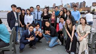
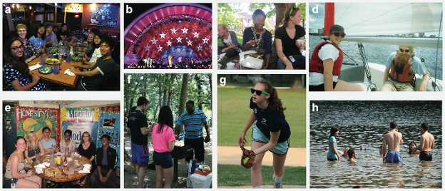
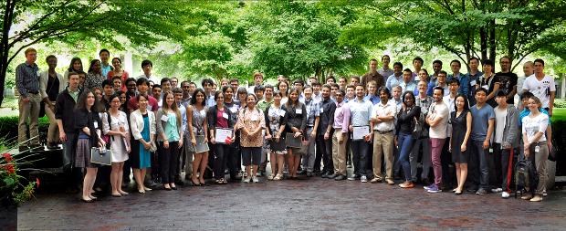
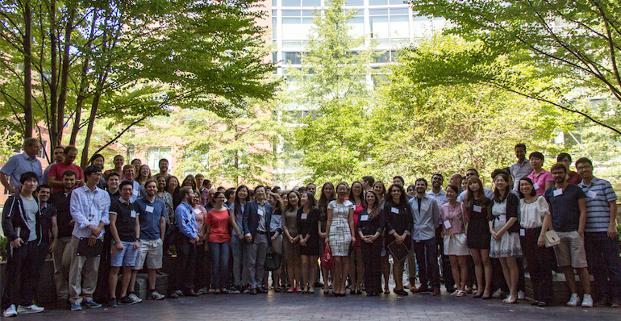
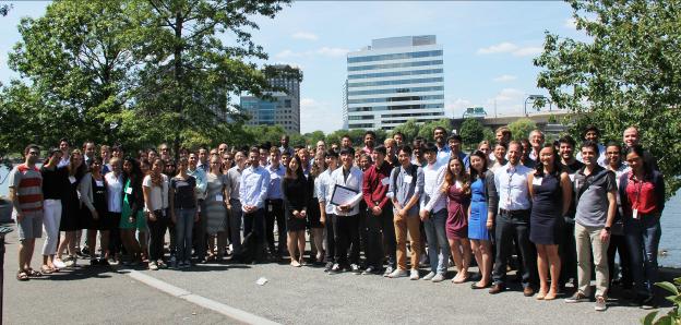
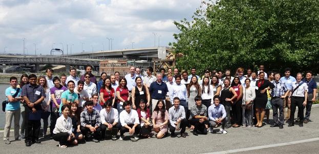
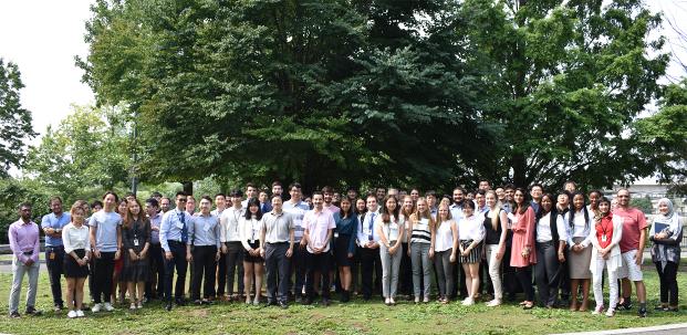
Community building and Social activities: Community-building activities play a central role in ensuring that students both enjoy their time here and develop friendships and acquaintances that will continue beyond the summer program. We take a proactive approach to building community over the short summer period. Summer program participants form a cohesive community that is strengthened by their close contact in a variety of settings.
We organize a variety of bi-weekly social activities to help students build a sense of community and to form close friendships (See pictures above). During the Orientation Day, we introduce mentors and staff members to the students and provide them with helpful materials, including a collection of tour guides and a list of various social and cultural events in the city of Boston and on the campuses of MIT and Harvard. Our official social program begins with ‘Dinner Out’ as an icebreaker during the first week. Other notable activities include a barbecue picnic in July with staff members and mentors, a Boston Pops Orchestra Concert on July 4th, and a softball game as a part of All-Wellman Summer Party. Independently, the students are encouraged to develop their own social activities during the weekends. Furthermore, interested students can arrange a visit to different labs in MIT and Harvard as well as to other schools in the Boston area. By the end of the summer, the participating students form close friendships, and many of former students maintain their contacts through Facebook.
Proposal writing and final report: In the third week of the program, after initial meetings with their research mentors, students develop a one-page research proposal for their summer project. The goal is to guide students in thinking through their research process so that they can reasonably articulate what they are going to do, how they intend to do it, why they are going to do it, what is likely to be concluded, and what additional work is necessary to conclude their projects. The proposal ensures that students and mentors establish a well-defined plan for the student’s summer research experience. In their proposals, students delineate a clear purpose and measurable aims for their research, possible alternative approaches, and expected results. Students write a five-page research report that includes a detailed explanation of their research goals, approach, and findings. Their faculty mentor and instructor review the draft and provide feedback and students submit their final reports in the final week.
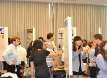
Poster preparation: To help the students improve their communications skills, we organize workshops on writing and research presentation. A group of experienced postdoctoral fellows and faculty members in collaboration with MIT’s Writing and Humanistic Studies Department provide two interactive sessions. Note that students submit a rough draft of their posters in PowerPoint format to the communication instructor and peers during a workshop. The group provides substantive feedback on the content, design, and delivery of the presentation. The goal is for students to articulate a coherent presentation, including the purpose of the project, the methods, the primary findings, a plausible interpretation of those findings, and the potential implications of the findings. In this process, students learn how to present their research findings in different formats using graphical, publishing, and presentation software.
Research presentation: In the final week, students present the results of their summer research to their faculty mentors, post-docs, and other students (see the photo). Each student gives a 10-minute poster presentation to invited judges. The judges select one student from each group who gives the best presentation for the Yao Su Student Research Award, which includes a certificate and $100 gift card from the endowment fund. After the poster session, the mentors award a certificate to their students in person.
Attending professional conferences Students are encouraged to present their reports at their home institution and submit posters to professional conferences. In the past, several students have presented their posters at the Annual Meeting of the Biomedical Engineering Society and SPIE BIOS meetings.
Student-and-faculty interactions: The variety of social and research events we organize throughout the summer ensures that the students and faculty build strong mentor-mentee relationships that can last even after the program ends. Our faculty mentors have maintained long-term relationships with many of their former students and, moreover, have provided recommendation letters for their applications to graduate schools, medical schools, scholarships, and beyond. In fact, 10 students have come back to WCP after receiving their undergraduate degrees as graduate students, postdocs, or research assistants. |


|
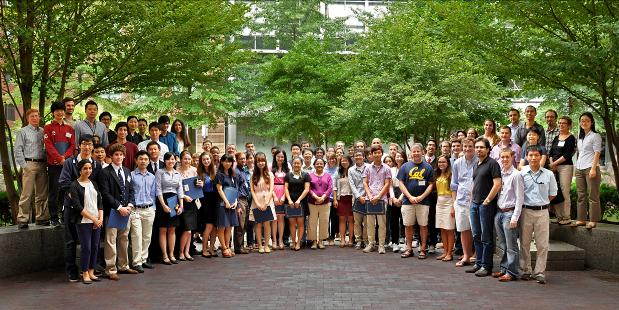
August 8, 2013 |
|
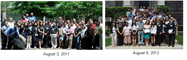
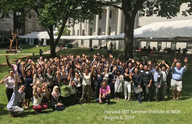
The Harvard-MIT Summer Institute at MGH |
|
August 7, 2015 |
|
August 7, 2014 |
|
August 5, 2016 |
|
August 4, 2017 |
|
August 17, 2018 |
|
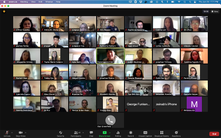
The 2021 Virtual Program (32 student participants from the U.S., Canada, and East Asia) |
|
August 8, 2019 |
|
The Summer REU Program (not offered in 2024) ————————————————————————————— Who is the program for? The full-time program is for undergraduate students within 1-2 years of graduation, who are interested in having hand-on experience on cutting-edge Biomedical Optics Research. Students from under-represented minority groups, first-generation college students, and students with disabilities are especially encouraged to apply. What can I expect? Full-time Mentored Research Experience: Each student will be assigned to a lab at Wellman Center and fully immersed in a student-owned project. Interactive Lessons Covering Aspects of Biomedical Optics: Learn about why light in medicine, fluorescence imaging, light-tissue interaction, photochemistry, and more, from experts Additional Benefits: • Stipend ($5000) • Travel expense support • Dorm • Network with students from across the US and internationally • Various fun activities including Red Sox game at Fenway • Compete for awards Key Dates: • Application deadline: closed • Admission finalized: ~by Feb • Program Duration: ~June 10 to August 9, 2024 • Certificate ceremony: August 8, 2024 Learn More: • Email: BioOpticsSummerInstitute@mgh.harvard.edu · See more details on the REU program toward the end of this page Application Form: Download from here PDF
————————————————————————————-
|
|
The 2020 program has been canceled due to COVID-19. |
|
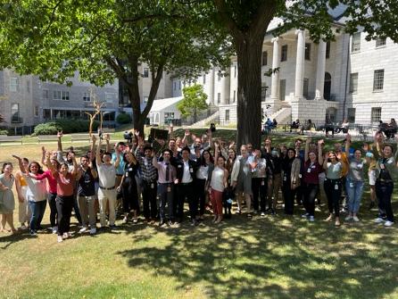
August 4, 2022 |
|
August 3, 2023 |